
Optics and Laser Technology 131 (2020) 106430

Contents lists available at ScienceDirect

Optics and Laser Technology
journal homepage: www.elsevier.com/locate/optlastec
Full length article
| Laser recording of color voxels in lithium fluoride | T |

E.F. Martynovicha,b, , E.O. Chernovab,c, V.P. Dresvyanskyb, A.E. Bugrovd, P.V. Kostryukovd, A.V. Konyashchenkod
- Irkutsk State University, Irkutsk 664003, Russia
- Irkutsk Branch of the Institute of Laser Physics of the SB RAS, Irkutsk 664033, Russia
- Peter the Great St. Petersburg Polytechnic University, St. Petersburg 195251, Russia
- P.N. Lebedev Physical Institute of the RAS, Moscow 119333, Russia
HIGHLIGHTS
- The laser beam creates colored voxels inside a transparent medium.
- Voxel colors are determined by their luminescence and light scattering spectra.
- Media with excitonic mechanism of defect creation are applicable for color recording.
- Recording technology includes three stages of laser irradiation and heat treatment.
ARTICLE INFO ABSTRACT
![]()
![]()
| Keywords: | The problem of creating three different types of spatially selective voxels by laser radiation in the volume of an |
| Laser | transparent optical medium was solved. Each of the created voxels provides one base color: red, green, or blue. |
| Femtosecond | Red and green voxels are microvolumes inside a crystal in which femtosecond laser pulses create luminescence |
| Crystal | centers that emit, respectively, in the red and green regions of the spectrum when illuminated with blue ra- |
| Color center | |
| diation that excites luminescence. Voxels responsible for the blue glow are microvolumes in which light scat- | |
| Voxel | |
| tering centers are created by femtosecond pulses. These voxels do not contain any luminescence centers; they | |
| Luminescence | |
| scatter blue radiation, which excites the luminescence of red and green voxels. To solve the problem of creating | |
| Spectrum | |
| spatially separated voxels of all three types in the volume of an optical carrier, we studied the temperature | |
| Excitation | |
| Image | dependences of the spectra, the output, and the decay time constants of the luminescence of quantum systems |
| Recording | created by femtosecond laser radiation. These dependences were studied during heating and cooling. The change |
| Lithium fluoride | in the voxel size of the three basic colors from the energy and the number of femtosecond pulses of a titanium- |
| sapphire laser was studied. Irradiation regimes were found in which only luminescence centers are formed in | |
| voxels and scattering centers are not formed. On the basis of the results, a three-stage technology of laser | |
| irradiation and heat treatment of optical media was proposed and experimentally tested. This technology allows | |
| for creation of three-dimensional, full-color images, visualized under blue light. | |
1. Introduction
Technologies for optical processing of materials are widely used to modify materials’ properties and to manufacture various functional elements of optical-electronic devices [1–7]. One of the current topical areas of research and technology is laser recording of digital and visual information in 3D and multi-layer optical media [8–19]. A promising direction of such studies is the formation of images visualized in their luminescent radiation [12–19]. In particular, in Refs. [17–19] lithium fluoride crystals were used as optical carrier, and color centers were
![]()
![]()
Corresponding author at: Irkutsk State University, Irkutsk 664003, Russia. E-mail address: femto@bk.ru (E.F. Martynovich).
used as work centers responsible for forming luminescent images. For the first time in the Refs. [20–25] it was argued that such luminescent color centers can be formed inside these crystals by intense femtose-cond laser radiation in the near IR region of the spectrum. Various properties of color centers created in crystals of lithium fluoride by hard radiation or by laser radiation, as well as some of their practical ap-plications, are described in Refs. [26–37]. In Ref. [17] it was proposed to use F2 color centers to form voxels responsible for the red color in images, and to use F3+ color centers for the green color in images. In the paper, the voxels responsible for the blue glow were represented as
https://doi.org/10.1016/j.optlastec.2020.106430
Received 14 August 2019; Received in revised form 13 May 2020; Accepted 13 June 2020
0030-3992/ © 2020 Elsevier Ltd. All rights reserved.
E.F. Martynovich, et al.
the points of laser destruction of the optical carrier material, scattering the blue radiation of the source used for luminescent visualization of the captured color images. However, the optimal method of recording the three types of such voxels was not proposed in Ref. [17]. In order to record an image inside the volume of the optical carrier, the laser ra-diation must freely pass to a given point in the medium without in-teracting with it. Conversely, at this point it is necessary to provide an effective highly nonlinear interaction that will lead to the formation of luminescent voxels. In lithium fluoride crystals, these conditions are automatically fulfilled for focused laser radiation in the visible and near-infrared regions of the spectra at a selected intensity value [18,36].
The processes of highly nonlinear internal photoionization and ex-citation occur under the action of laser radiation in a lithium fluoride crystal. Anionic excitons, which arise upon recombination of charge carriers and upon their nonlinear excitation, decay into anionic Frenkel defects. In parallel, processes of recharging and migration of defects occur. After laser irradiation, aggregation processes lead to the forma-tion of the first stable aggregate centers F2 and F3+, which have, re-spectively, red and green photoluminescence, characterized by a high yield [38,39].
To generate color images the voxels of the three base colors need to be spatially separated. However, F2 and F3+ centers in lithium fluoride crystals irradiated with laser radiation or hard radiation are always formed together. In addition, they have completely overlapping ab-sorption bands. This presents the main problem – creating spatially separated monochrome voxels of different colors in the same optical medium. Overcoming this problem was the subject of our work. For this purpose, the mechanisms of formation of the color centers and their further transformation under external impact were considered. In par-ticular, temperature dependences of the intensity and the luminescence decay time constants of separate types of color centers, as well as their thermal stability, were researched. The obtained results allowed to re-cord spatially separated voxels of the three base colors in the same block of an optical carrier. This made it possible to justify the tech-nology of recording of 3D, full-color images in optical media based on lithium fluoride crystals.
2. Methods
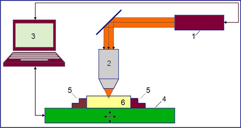
The technology for laser recording of volumetric color images inside an optical carrier based on a lithium fluoride crystal, proposed in this article, can be implemented using the technological setup shown in Fig. 1. Preliminary experiments showed that the voxels of the three types described above can be created by various femtosecond lasers in
Optics and Laser Technology 131 (2020) 106430
the near-IR spectral region with a pulse energy of less than 1 mJ. In this paper, we present the results obtained using titanium-sapphire laser complexes created by Avesta-Project [40]. Laser irradiation of crystals was carried out at the facilities of the Physical Institute of the Russian Academy of Sciences and the Institute of High Current Electronics of the SB RAS. In order to determine the types of color centers created by radiation in crystals, we studied the absorption and luminescence spectra of irradiated samples, as well as the decay time of luminescence. The absorption spectra are conveniently measured on samples with a relatively large irradiated area. For such measurements, crystal plates with a thickness of ~3 mm were irradiated. 5 × 5 mm2 sites were irradiated on these samples in the line scan mode using a lens with a focal length of 40 cm. The absorption spectra of induced color centers were measured with an SF-56 spectrophotometer. The luminescence spectra and its decay time were studied using a confocal scanning lu-minescent microscope with a time resolution MicroTime 200 equipped with an Ocean Optics 65,000 spectrofluorimeter. The thermal de-struction and transformation of color centers, as well as the tempera-ture dependences of the luminescence intensity and its decay time, were studied using a Microstat-N cryostat from Oxford Instruments, or a special external heater.
In addition to the described measurements, studies of the sizes of single voxels of all three types were carried out depending on the conditions and modes of irradiation. The radiation of femtosecond laser pulses was focused with a 40x / 0.60 planachromat objective at a depth of 200 μm from the crystal surface to form voxels. Other experimental conditions and modes are listed below.
3. Results and discussion
The measured absorption spectrum in Fig. 2 indicats that the main centers formed when being laser-irradiated are F centers (intense shortwave band in the absorption spectrum), as well as F2 and F3+ centers which form a complex absorption band with a maximum at 443 nm. In the lithium fluoride crystal F centers do not luminesce. The luminescence spectra excited in the second induced absorption band, contain two known spectral bands corresponding to the F2 (670 nm) and F3+ centres (540 nm) [41,42]. The measured values of the lumi-nescence decay time constants in these bands of 16 and 7–8 ns (as presented below in Fig. 5b) confirm that these bands correspond to the described centers.
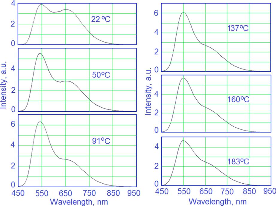
The results in Fig. 3 show that the contribution of these two centers in the overall intensity of luminescence of the irradiated sites varies depending on the temperature. One can notice that the intensity of luminescence F3+ with rise in temperature from 22 up to 183 °C grows,
Fig. 1. Experimental setup: 1 – femtosecond laser, 2 – focusing system; 3 – computer; 4 – movable positioner table; 5 – clamp; 6 – optical carrier.
2
E.F. Martynovich, et al. Optics and Laser Technology 131 (2020) 106430
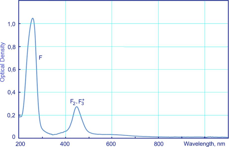
Fig. 2. Optical absorption spectrum of an irradiated crystal.
reaches a maximum at the temperature of 91 °C and then decreases. Within this temperature range, the intensity of luminescence in F2 centers decreases slightly. As a result, the ratio of luminescence in-tensities of these centers is changing in favor of F3+ centers luminescing in the green region of the spectrum.
With temperature rising, there is an accelerated reduction in lumi-nescence intensity of F3+ centers. This follows from the data in Fig. 4, which shows the luminescence spectra in the range of 270–300 °C. It can be seen that at temperatures above 283 °C, the only remaining luminescence band in the spectrum is that of F2 centers, which are re-sponsible for voxels of red.
The intensity or emission of luminescence can depend on tem-perature due to two reasons: 1 – intracenter luminescence quenching due to nonradiative transitions and 2 – thermal conversion or
destruction of the centers responsible for luminescence. If the tem-perature dependences are due to intracenter quenching, and the prob-ability of nonradiative transitions b is determined by the Mott formula [43],
| ΔE | , | ||
| b = b0exp − | kT | ||
| (1) | |||
then the luminescence yield η and the decay time constant τ change with temperature in accordance with the expressions:
| η = | a | ||
| a + b0 exp( − | ΔkTE ) | (2) | |
![]()
![]()
Fig. 3. Luminescence spectra recorded at different temperatures during the heating of the sample at the same sensitivity of the setup (λexc = 450 nm).
3
E.F. Martynovich, et al. Optics and Laser Technology 131 (2020) 106430

Fig. 4. Luminescence spectra measured at temperatures indicated in the figures during heating. The lowest curve shows the spectrum of a crystal heated at 300 °C, measured at room temperature 23 °C after cooling. The sensitivity of the experimental setup is the same as in Fig. 3 (λexc = 450 nm).
| τ = | 1 | , | |
| a + b0 exp( −ΔkTE ) | (3) |
where a – radiative transition probability, bo – frequency factor, ΔE – quenching activation energy, k – the Boltzmann constant, T – the ab-solute temperature.
It follows from the above expressions (2) and (3) that the tem-perature dependences of the luminescence yield and luminescence decay time constant are identical if they are due to nonradiative in-tracenter transitions, but not to thermal changes of the concentrations of centers. To answer the question of the presence of intracenter quenching, the measured temperature intensity dependences for each center were compared with the relevant experimentally measured temperature dependences of the decay time constants. To examine the contribution to the observed temperature dependences of the thermal conversion or destruction of the color centers, the luminescence in-tensities of some color centers were measured both during heating and cooling of a crystal. These data are shown in the Fig. 5.
From these data, it follows that for F3+ centers the temperature dependences of luminescence intensity and decay time constant mea-sured during heating vary considerably. However, on cooling, they are almost identical. The same is observed for F2 centers. Small deviations are caused by the temperature dependences of the shapes of the ex-citation and luminescence bands of these centers. In light of the results obtained, we conclude that: 1 – temperature luminescence quenching of both types of centers in the 23–300 °C range is insignificant, 2 – a de-crease in the luminescence intensity of F3+ and F2 centers upon heating of the crystal is due to a decrease in their concentrations, 3 – consistent increase and decrease in the luminescence intensities of the centers F3+
- F2 in the temperature range of 20–100 °C are due to the corre-sponding changes in the concentrations of these centers. This concerted process is driven by the thermal splitting of anionic vacancies Va+ from radiation defects of unknown origin, their migration and subsequent aggregation with an F2 centers in the following reaction:
F2 + V a+ → F3+.
As can be seen from this reaction, in the course of the process, the concentration of F2 centers drops while the concentration of F3+ cen-ters increases, which is observed in the experiment.
Thus, thermal processing of irradiated crystals can be used to
Fig. 5. Temperature dependences of luminescence intensity spectra integrated F2 and F3+ centers (a) and their decay time constants (b). The arrows indicate the direction of temperature change.
optimize the spectra of voxels responsible for the green and red ra-diation.
The data obtained made it possible to formulate and recommend a sequence of three stages of laser irradiation and heat treatment to form three types of spatially separated voxels, providing one of the three basic colors each, in the same optical medium based on a lithium fluoride crystal.
The first step is to create light-scattering voxels responsible for the blue color of the image. At this stage it is necessary to irradiate the media by laser radiation at room temperature. The energy and the number of pulses of femtosecond radiation must be selected in such a way as to form the points of destruction of the carrier material (see below). At the same time, under the action of laser irradiation, lumi-nescent color centers are formed together with light scattering voxels. To eliminate them, the optical carrier must be removed from the po-sitioner’s movable stage, placed in the heater, and kept at a temperature above 400 °C until the F2 and F3+ color centers are completely de-stroyed. A trial experiment showed that exposure at this temperature for 5 min leads to the complete destruction of these centers. These data on the thermal stability of color centers are consistent with the results of Ref. [44].
At the second stage, the optical medium 6 containing the formed blue voxels must be reinstalled on the movable table of the positioner 4, centered and fixed using the latch 5. At this stage, voxels responsible for the red color of the image are created in the optical medium. For this, the carrier must be irradiated with femtosecond laser radiation. In this case, two types of luminescent centers F2 and F3+ with red and green luminescence, respectively, are formed at the irradiated points. The irradiation regimes must be chosen so as to prevent laser destruction of the carrier material, leading to light scattering (see below). To destroy F3+ centers, the crystal must be removed from the table, heated and held at temperatures in the range from 270 to 300 °C. The color centers remaining after this F2 treatment provide the red color of the resulting voxels. The luminescence spectrum of these voxels is shown by the lower curve in Fig. 4.
The third stage provides for the creation of voxels responsible for the green color of the image. The media is to be irradiated by femto-second laser radiation, heated to a temperature in the 50–160 °C range,
4
E.F. Martynovich, et al. Optics and Laser Technology 131 (2020) 106430
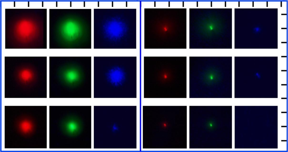
FIG. 6. Change in the size of voxels when changing the number of radiation pulses of a titanium-sapphire laser (950 nm, 50 ns) and their energy: on the left, the pulse energy is 9 μJ; on the right, −1.5 μJ; the top row − 1350 pulses, the middle row − 50 pulses (pulse repetition rate 10 Hz), the bottom row – one pulse. When photographing voxels, they were visualized by continuous radiation of a laser diode with a wavelength of 450 nm. Scale − 20 µm / division. Images taken on an Inverted microscope Olympus IX 73.
and kept at that temperature to the maximum concentration of F3+ centers. As follows from the data given in Figs. 3 and 4, the optimum temperature is around 100° C. The luminescence spectra of voxels after such heat treatment are shown in Fig. 3 contain a noticeable con-tribution of the red band from the residual F2 centers. It is not possible to completely get rid of them. The color of such voxels is visually perceived as yellow-green.
It is important to emphasize that each subsequent stage of the op-tical media processing listed above does not change the properties of the voxels generated at the previous stages.
To visualize the recorded image, its luminescence is excited by ra-diation of the blue region of the spectrum. This allows us to visualize the voxels responsible for the red and green glow. The third base color is reproduced by light scattering pixels, which are colored blue, scat-tering radiation that excites luminescence.
In Fig. 6 shows photographs of individual voxels of the three de-scribed types formed in an optical medium under various laser irra-diation conditions. It is seen that the size of the voxels increases sig-nificantly with increasing both the energy of the pulses and their number. Pulses with an energy of 1.5 μJ do not yet lead to the de-struction of the crystalline medium; therefore, they do not form light scattering voxels responsible for the blue color. At the same time, they successfully create color centers that provide the formation of red and green voxels. Single impulses with an energy of 9 μJ already confidently produce destruction in the bulk of the crystal. Therefore, they can be used to form blue voxels. It is noteworthy that a series of 50 and 1350 pulses with an energy of 1.5 μJ also produces light scattering voxels (see Fig. 7).
4. Conclusion
Thus, in this work, a physical justification is given for the tech-nology for recording color 3D images in a transparent optical carrier based on a lithium fluoride crystal. This technology is based on the found possibilities of creating voxels of three basic colors: red, green and blue under the influence of laser radiation. The technology includes three stages of femtosecond laser irradiation and three stages of heat
treatment of the optical medium. Voxels that provide red and green colors of the image contain, respectively, F2 and F3+ color centers. These centers luminesce in the red and green regions of the spectrum when they are excited by blue light. Blue voxels are microregions of laser destruction of a lithium fluoride crystal that do not contain lu-minescent color centers and scatter the blue light of a source that ex-cites the luminescence of green and red voxels.
It was shown that, using a 40x/0.60 focusing lens, single pulses of a titanium-sapphire laser (950 nm, 50 fs) with an energy of less than 1.5 μJ form luminescent color centers in the crystal, but do not lead to destruction of the material, which causes light scattering. Therefore, such pulses can be used to record red and green voxels. On the other hand, for the formation of blue voxels with single laser pulses, it is necessary to select an energy of more than 1.5 μJ. There are also pos-sibilities to control the size of voxels by changing the energy and number of pulses of laser irradiation.
CRediT authorship contribution statement
E.F. Martynovich: Conceptualization, Methodology, Validation, Formal analysis, Investigation, Resources, Writing – original draft, Writing – review & editing, Supervision, Project administration, Funding acquisition. E.O. Chernova: Validation, Investigation, Writing
- original draft, Writing – review & editing, Visualization. V.P. Dresvyansky: Methodology, Validation, Formal analysis, Investigation, Resources. A.E. Bugrov: Methodology, Validation, Software, Formal analysis, Investigation, Resources. P.V. Kostryukov: Methodology, Validation, Software, Formal analysis, Investigation, Resources. A.V. Konyashchenko: Methodology, Validation, Software, Formal analysis, Investigation, Resources.
Declaration of Competing Interest
The authors declare that they have no known competing financial interests or personal relationships that could have appeared to influ-ence the work reported in this paper.
5
E.F. Martynovich, et al. Optics and Laser Technology 131 (2020) 106430
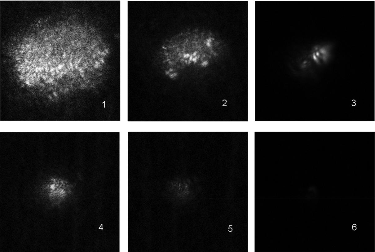
Fig. 7. Scanned images of light scattering voxels created at a pulse energy of 9 μJ (upper row) and 1.5 μJ (lower row). Scanning was performed by picosecond pulses with a wavelength of 470 nm. Voxels 1 and 4 are created by irradiation with a series of 1350 pulses, 2 and 5 – from 50 pulses, 3 and 6 – single pulses. The frequency in the series is 10 Hz. Images were obtained using a MicroTime200 confocal scanning microscope. 40x lens. Image size 30×30 μm2.
Acknowledgement
Corresponding author expresses sincere gratitude to Academician A.N. Ratakhin and Prof. V.F. Losev for the opportunity to irradiate crystals with a femtosecond laser at the Institute of High Current Electronics SB RAS, S.V. Alekseev for help in irradiating, and A.L. Rakevich for helping with experiments using the MicroTime 200 mi-croscope .
This work was supported by State Task of the Ministry of Education and Science of the Russian Federation for Implementation of Scientific Research (the basic part), Project No. 3.8401-2017/8.9 and by the Program of Fundamental Scientific Research of the State Academies of Sciences of the Russian Federation for 2013–2020, section II.10.1 Project No. 0307-2016-0004.
References
- Serguei P. Murzin, Gerhard Liedl, Robert Pospichal, Coloration of a copper surface by nanostructuring with femtosecond laser pulses, Opt. Laser Technol. 119 (2019) 105574, https://doi.org/10.1016/j.optlastec.2019.105574.
- J. Li, D.u. Feng, Y. Zhao, S. Zhao, Y.u. Huadong, Two–step fabrication of super-hydrophobic surfaces with anti–adhesion, Opt. Laser Technol. 113 (2019) 273–280, https://doi.org/10.1016/j.optlastec.2018.12.045.
- J. Hernandez-Rueda, J. Clarijs, D. van Oosten, D.M. Krol, The influence of femto-second laser wavelength on waveguide fabrication inside fused silica, Appl. Phys. Lett. 110 (2017) 161109, https://doi.org/10.1063/1.4981124.
- X. Wang, A.A. Kuchmizhak, E. Brasselet, S. Juodkazis, Dielectric geometric phase optical elements fabricated by femtosecond direct laser writing in photoresists, Appl. Phys. Lett. 110 (2017) 181101, https://doi.org/10.1063/1.4982602.
- L. Yang, D. Qian, C. Xin, Hu Zhijiang, Shengyun Ji, Wu Dong, Hu Yanlei, Jiawen Li, Wenhao Huang, Jiaru, Direct laser writing of complex microtubes using femtose-cond vortex beams, Appl. Phys. Lett. 110 (2017) 221103, https://doi.org/10.1063/ 1.4984744.
- R. Osellame, M. Lobino, N. Chiodo, M. Marangoni, G. Cerullo, R. Ramponi, H.T. Bookey, R.R. Thomson, N.D. Psaila, A.K. Kar, Femtosecond laser writing of waveguides in periodically poled lithium niobate preserving the nonlinear
coefficient, Appl. Phys. Lett. 90 (2007) 241107, https://doi.org/10.1063/1.
- A. Solodar, A. Cerkauskaite, R. Drevinskas, P.G. Kazansky, I. Abdulhalim, Ultrafast laser induced nanostructured ITO for liquid crystal alignment and higher trans-parency electrodes, Appl. Phys. Lett. 113 (2018) 081603, https://doi.org/10.1063/ 1.5040692.
- R.R. Gattass, E. Mazur, Femtosecond laser micromachining in transparent materials, Nat. Photon. 2 (2008) 219, https://doi.org/10.1038/nphoton.2008.47.
- J. Zhang, A. Čerkauskaitė, R. Drevinskas, A. Patel, M. Beresna, P.G. Kazansky, Eternal 5D data storage by ultrafast laser writing in glass, in: Proc. SPIE 9736, Laser-based Micro- and Nanoprocessing X, 97360U, 2016. https://doi.org/10.1117/12. 2220600.
- R. Drevinskas, P.G. Kazansky, Seemingly unlimited lifetime data storage in nanos-tructured glass, Phys. Rev. Lett. 112 (2014) 033901, https://doi.org/10.1103/ PhysRevLett.112.033901.
- R. Drevinskas, P.G. Kazansky, High-performance geometric phase elements in silica glass, APL Photon. 2 (2017) 066104, https://doi.org/10.1063/1.4984066.
- N. Marquestaut, Y. Petit, A. Royon, P. Mounaix, T. Cardinal, L. Canioni, Three-dimensional silver nanoparticle formation using femtosecond laser irradiation in phosphate glasses: analogy with photography, Adv. Funct. Mater. 24 (37) (2014) 5824–5832, https://doi.org/10.1002/adfm.201401103.
- C. Maurel, T. Cardinal, M. Bellec, L. Canioni, B. Bousquet, M. Treguer, J.J. Videau,
- Choi, M. Richardson, Luminescence properties of silver zinc phosphate glasses following different irradiations, J. Lumin. 129 (12) (2009) 1514–1518, https://doi. org/10.1016/j.jlumin.2008.12.023.
- M. Vangheluwe, Y. Petit, N. Marquestaut, A. Corcoran, E. Fargin, R. Vallée,
- Canioni, Nanoparticle generation inside Ag-doped LBG glass by femtosecond laser irradiation, Opt. Mater. Express 6 (3) (2016) 743–748, https://doi.org/10. 1364/OME.6.000743.
- A. Royon, K. Bourhis, M. Bellec, G. Papon, B. Bousquet, Y. Deshayes, T. Cardinal,
- Canioni, Silver clusters embedded in glass as a perennial high capacity optical recording medium, Adv. Mater. 22 (2010) 5282–5286, https://doi.org/10.1002/ adma.201002413.
- E. Lee, Y. Petit, E. Brasselet, T. Cardinal, S.H. Park, L. Canioni, Sub-diffraction-limited fluorescent patterns by tightly focusing polarized femtosecond vortex beams in a silver-containing glass, Opt. Express 25 (9) (2017) 10565–10573, https://doi. org/10.1364/OE.25.010565.
- A.V. Kuznetsov, L.I. Bryukvina, E.F. Martynovich, 3D image media (Nositel’ ob“yemnogo izobrazheniya), Patent RU 135964 U1. Bulletin # 36, 2013. (in Russian). http://www.fips.ru/cdfi/fips.dll/ru?ty=29&docid=135964&ki=PM.
- E.F. Martynovich, A.V. Kuznetsov, A.V. Kirpichnikov, E.V. Pestryakov,
6
E.F. Martynovich, et al.
S.N. Bagayev, Formation of luminescent emitters by intense laser radiation in transparent media, Quantum Electron. 43 (5) (2013) 463–466, https://doi.org/10. 1070/QE2013v043n05ABEH015117.
- E.F. Martynovich, V.P. Dresvyansky, N.L. Lazareva, S.V. Mikhailova,
A.V. Konyashchenko, P.V. Kostryukov, B.E. Perminov, S.N. Bagayev, The memor-izing luminescent crystalline materials based on color centers for investigation the highly nonlinear interaction of light and matter and for other applications, Adv.
Photonics, OSA, NoW2C.6 (2017), https://doi.org/10.1364/NOMA.2017. NoW2C.6.
- T. Kurobori, K. Kawamura, M. Hirano, H. Hosono, Simultaneous fabrication of laser-active colour centres and permanent microgratings in lithium fluoride by a single femtosecond pulse, J. Phys: Cond. Matter 15 (2003) L399–L405, https://doi. org/10.1088/0953-8984/15/25/101.
- K. Kawamura, M. Hirano, T. Kurobori, D. Takamizu, T. Kamiya, H. Hosono, Femtosecond-laser-encoded distributed-feedback color center laser in lithium fluoride single crystals, Appl. Phys. Lett. 84 (2004) 311–313, https://doi.org/10. 1063/1.1640784.
- T. Kurobori, T. Yamakage, Y. Hirose, K. Kawamura, M. Hirano, H. Hosono, Applications of wide-band-gap materials for optoelectronic functional devices fab-ricated by a pair of interfering femtosecond laser pulses, Jpn. J. Appl. Phys. 44 (2005) 910–913, https://doi.org/10.1143/JJAP.44.910.
- L.C. Courrol, R.E. Samad, L. Gomes, I.M. Ranieri, S.L. Baldochi, A. Zanardi de Freitas, N.D. Vieira, Color center production by femtosecond pulse laser irradiation in LiF crystals, Opt. Express 12 (2) (2004) 288–293, https://doi.org/10.1364/ OPEX.12.000288.
- L.C. Courrol, R.E. Samad, L. Gomes, I.M. Ranieri, S.L. Baldochi, A.Z. Freitas, S.P. Morato, N.D. Vieira Jr., Color center production by femtosecond-pulse laser irradiation in fluoride crystals, Laser Phys. 16 (2) (2006) 331–335, https://doi.org/ 10.1134/S1054660X06020216.
- L.C. Courrol, R.E. Samad, O.E. Martinez, L. Gomes, I.M. Ranieri, S.L. Baldochi, A.Z. de Freitas, N.D. Vieira Jr., Determination of a dose-like curve for active colour centres produced in LiF single crystals by ultrashort high intensity laser pulses and a preliminary investigation of their spectral and spatial properties by confocal and atomic microscopies, J. Opt. A: Pure Appl. Opt. 10 (10) (2008), https://doi.org/10. 1088/1464-4258/10/10/104023.
- N.W. Jenkins, S.B. Mirov, V.V. Fedorov, Temperature-dependent spectroscopic analysis of F2+** and F2+**-like color centers in LiF, J. Lumin. 91 (2000) 147, https://doi.org/10.1016/S0022-2313(00)00228-3.
- V.V. Fedorov, S.B. Mirov, M. Ashenafi, L. Xie, Spectroscopic analysis and persistent photon-gated spectral hole burning in LiF:F2– – color center crystal, Appl. Phys. Lett. 79 (2001) 2318, https://doi.org/10.1063/1.1406563.
- R.M. Montereali, M. Piccinini, E. Burattini, Amplified spontaneous emission in ac-tive channel waveguides produced by electron-beam lithography in LiF crystals, Appl. Phys. Lett. 78 (2001) 4082, https://doi.org/10.1063/1.1381568.
- G. Tomassetti, A. Ritucci, A. Reale, L. Arrizza, F. Flora, R.M. Montereali, A. Faenov, T. Pikuz, Two-beam interferometric encoding of photoluminescent gratings in LiF crystals by high-brightness tabletop soft x-ray laser, Appl. Phys. Lett. 85 (2004)
4163, https://doi.org/10.1063/1.1812841.
- E.F. Martynovich. Color centers in laser crystals (Tsentry okraski v lazernykh kristallakh). Irkutsk: Irkutsk State University Press, 2004, 226 pp. (in Russian).
Optics and Laser Technology 131 (2020) 106430
- L.A. Lisitsyna, V.M. Lisitsyn, Composition nanodefects in doped lithium fluoride crystals, Phys. Solid State 55 (11) (2013) 2297, https://doi.org/10.1134/ S1063783413110139.
- L.A. Lisitsyna, R. Kassymkanova, V.M. Lisitsyn, Features of F2 centers accumulation in oxygen-containing LiF crystals, Adv. Mater. Res. 880 (2014) 62–67, https://doi. org/10.4028/www.scientific.net/AMR.880.62.
- E.F. Martynovich, V.P. Dresviansky, A.V. Kuznetsov, A.S. Kuzakov, A.A. Popov, S.V. Alekseev, V.F. Losev, A.N. Ratakhin, S.N. Bagayev, Simulation of filamentation of single femtosecond laser pulses in LiF, Laser Phys. 24 (7) (2014) 074001, https:// doi.org/10.1088/1054-660X/24/7/074001.
- E.F. Martynovich, V.P. Dresvyansky, A.L. Rakevich, N.L. Lazareva, M.A. Arsentieva, A.A. Tyutrin, O. Bukhtsoozh, S. Enkhbat, P.V. Kostryukov, B.E. Perminov,
A.V. Konyashchenko, Creating of luminescent defects in crystalline media by a scanning laser beam, Appl. Phys. Lett. 114 (2019) 121901, https://doi.org/10. 1063/1.5087688.
- A.P. Voitovich, V.S. Kalinov, V.V. Mashko, A.N. Novikov, L.P. Runets, A.P. Stupak, Transformation and formation of radiation-induced point defects in irradiated li-thium fluoride crystals after their mechanical fragmentation, J. Appl. Spectrosc. 86
(1) (2019) 61–66, https://doi.org/10.1007/s10812-019-00781-w.
- E.F. Martynovich, D.S. Glazunov, A.V. Kuznetsov, E.V. Pestriakov, A.V. Kirpichnikov, S.N. Bagayev, 3D Fluorescent Imaging with Highly Nonlinear Photosensitive Materials, Advanced Photonics, OSA, paper SWD6, 2011. https:// doi.org/10.1364/SENSORS.2011.SWD6.
- A.V. Kuznetsov, V.O. Kompanets, A.E. Dormidonov, S.V. Chekalin, S.A. Shlenov, V.P. Kandidov, Periodic colour-centre structure formed under filamentation of mid-IR femtosecond laser radiation in a LiF crystal, Quantum Electron. 46 (4) (2016) 379–386, https://doi.org/10.1070/QEL16038.
- E.F. Martynovich, D.S. Glazunov, A.A. Grigorova, A.A. Starchenko,
A.M. Kirpichnikov, V.I. Trunov, M.A. Merzlyakov, V.V. Petrov, E.V. Pestryakov, Highly nonlinear fundamental mechanisms of excitation and coloring of wide-gap crystals by intense femtosecond laser pulses, Opt. Spectrosc. 105 (3) (2008)
348–351, https://doi.org/10.1134/S0030400X0809004X.
- E.F. Martynovich, D.V. Balyunov, A.V. Kuznetsov, A.V. Kirpichnikov, V.I. Trunov, E.V. Pestryakov, S.N. Bagaev, Coloration of wide-gap crystals with intense laser radiation. Izvestiya vysshikh uchebnykh zavedeniy, Fizika 52 (2–3) (2009) 191–197 https://elibrary.ru/item.asp?id=24340123.
- http://avesta.ru/product/reus-usilitelnye-femtosekundnye-sistemy-na-titan-sapfire/.
- J. Nahum, D.A. Weigand, Optical properties of some F-aggregate centers in LiF, Phys. Rev. 154 (3) (1967) 817–830, https://doi.org/10.1103/PhysRev.154.817.
- J. Nahum, Optical properties and mechanism of formation of some F-aggregate centers in LiF, Phys. Rev. 158 (3) (1967) 814–825, https://doi.org/10.1103/ PhysRev.158.814.
- N.F. Mott, On the absorption of light by crystals, Proc. Royal Soc. A 167 (930) (1938) 384–391, https://doi.org/10.1098/rspa.1938.0137.
- A. Lushchik, I. Kudryavtseva, P. Liblik, Ch Lushchik, A.I. Nepomnyashchikh,
K. Schwartz, E. Vasil‘chenko, Electronic and ionic processes in LiF: Mg, Ti and LiF single crystals, Radiat. Measur. 43 (2–6) (2008) 157–161, https://doi.org/10.1016/ j.radmeas.2007.10.001.